Clinical Applications
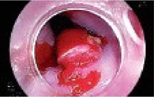
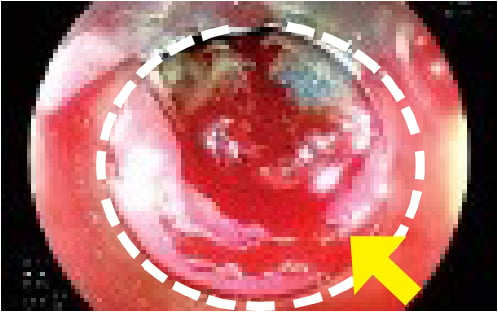
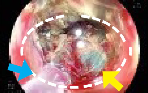
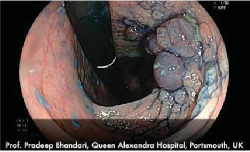
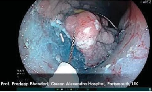
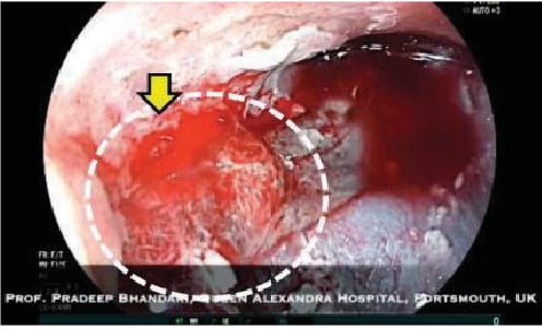
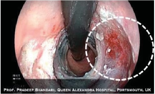
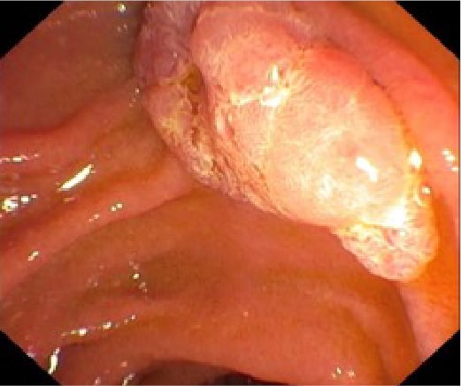
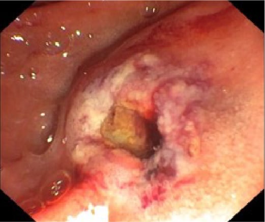
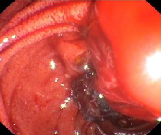
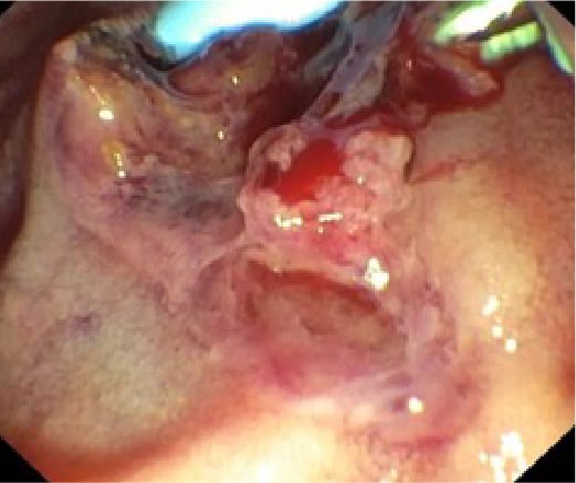
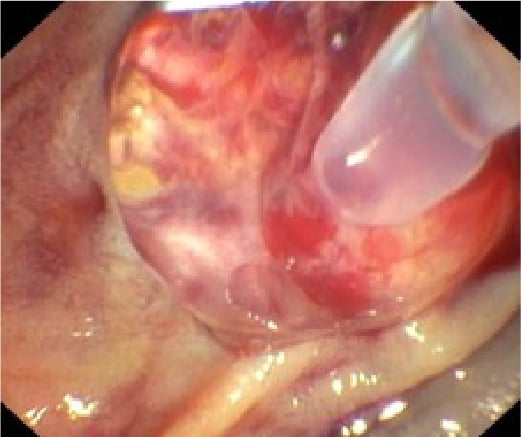
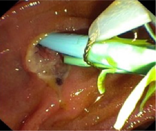
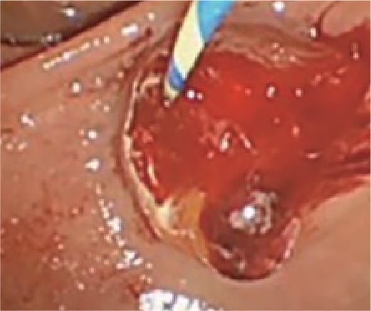
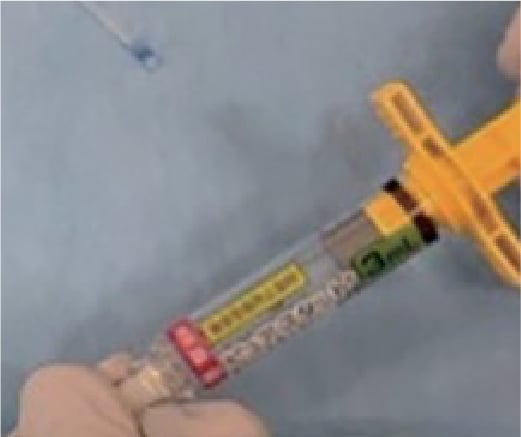
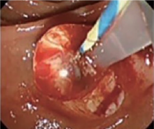
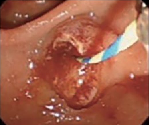
PuraStat is intended for hemostasis of mild and moderate bleeding post ESD or EMR, as an adjunct, bridge, prophylactic or rescue therapy for intraprocedural venous bleeding or prophylactic therapy to prevent post procedure bleeding in Gastrointestinal (GI) bleeding.
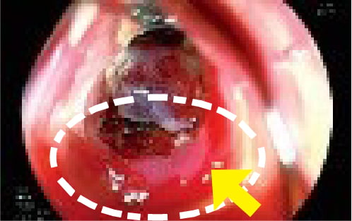
PuraStat is also indicated for the symptomatic management of Rectal Mucositis (RM), such as radiation proctitis that may be caused by chemotherapy or radiotherapy. Click tabs below for examples.
Resection
See how these examples demonstrate the application of PuraStat during resection procedures, such as ESD, EMR, and polypectomy.
Hemostasis with PuraStat ® During ESD/EMR Procedure
![Fig. 1: Barrett’s intramucosal cancer](https://3dmatrix.com/hs-fs/hubfs/3dmatrix_May2024/images/hemo1.jpg?width=495&height=312&name=hemo1.jpg)
Fig. 1: Barrett’s intramucosal cancer
![Fig. 2: Bleeding during ESD. Yellow arrow indicates bleeding point; White dot circle indicates resected area](https://3dmatrix.com/hs-fs/hubfs/3dmatrix_May2024/images/hemo2.jpg?width=498&height=312&name=hemo2.jpg)
Fig. 2: Bleeding during ESD. Yellow arrow indicates bleeding point; White dot circle indicates resected area
![Fig. 3: White dot circle: transparent PuraStat covered the bleeding site: Yellow arrow: bleeding point; Blue arrow: tip of the catheter](https://3dmatrix.com/hs-fs/hubfs/3dmatrix_May2024/images/hemo3.jpg?width=496&height=313&name=hemo3.jpg)
Fig. 3: White dot circle: transparent PuraStat covered the bleeding site: Yellow arrow: bleeding point; Blue arrow: tip of the catheter
![Fig. 4: Hemostasis achieved](https://3dmatrix.com/hs-fs/hubfs/3dmatrix_May2024/images/hemo4.jpg?width=498&height=313&name=hemo4.jpg)
Fig. 4: Hemostasis achieved
Source 1–4: Professor Pradeep Bhandari, Queen Alexandra Hospital, United Kingdom
Resection of a Large Circumferential, Recto-sigmoid Polyp
![Fig. 1: Carpet adenoma of rectum](https://3dmatrix.com/hs-fs/hubfs/3dmatrix_May2024/images/res1.jpg?width=495&height=301&name=res1.jpg)
Fig. 1: Carpet adenoma of rectum
![Fig. 2: Snare resection of the lesion](https://3dmatrix.com/hs-fs/hubfs/3dmatrix_May2024/images/res2.jpg?width=495&height=301&name=res2.jpg)
Fig. 2: Snare resection of the lesion
![Fig. 3: White dot circle: transparent PuraStat covered the bleeding site; Yellow arrow: bleeding point](https://3dmatrix.com/hs-fs/hubfs/3dmatrix_May2024/images/res3.jpg?width=496&height=301&name=res3.jpg)
Fig. 3: White dot circle: transparent PuraStat covered the bleeding site; Yellow arrow: bleeding point
![Fig. 4: Hemostasis was achieved](https://3dmatrix.com/hs-fs/hubfs/3dmatrix_May2024/images/res4.jpg?width=495&height=301&name=res4.jpg)
Fig. 4: Hemostasis was achieved
Source 1–4: Professor Pradeep Bhandari, Queen Alexandra Hospital, United Kingdom
Using PuraStat to Manage Bleeding in Endoscopic Resection (Professor Bhandari)
Using PuraStat during Cecal Polyp Resection (Professor Neumann)
ERCP
See how these examples demonstrate the application of PuraStat during procedures in conjunction with ERCP.
Hemostasis with PuraStat ® in Papillectomy
![Fig.1: Adenoma of the duodenal major papilla](https://3dmatrix.com/hs-fs/hubfs/3dmatrix_May2024/images/hemo1-2.jpg?width=521&height=437&name=hemo1-2.jpg)
Fig.1: Adenoma of the duodenal major papilla
![Fig. 2: Unremarkable resection site after endoscopic papillectomy](https://3dmatrix.com/hs-fs/hubfs/3dmatrix_May2024/images/hemo2-2.jpg?width=521&height=437&name=hemo2-2.jpg)
Fig. 2: Unremarkable resection site after endoscopic papillectomy
![Fig. 3: Severe arterial bleeding on the following day](https://3dmatrix.com/hs-fs/hubfs/3dmatrix_May2024/images/hemo3-2.jpg?width=521&height=437&name=hemo3-2.jpg)
Fig. 3: Severe arterial bleeding on the following day
![Fig. 4: Persistent bleeding despite of injection of adrenaline (1:10000)](https://3dmatrix.com/hs-fs/hubfs/3dmatrix_May2024/images/hemo4-2.jpg?width=521&height=437&name=hemo4-2.jpg)
Fig. 4: Persistent bleeding despite of injection of adrenaline (1:10000)
![Fig. 5: Transparent PuraStat covered the bleeding site. Complete hemostasis was achieved after 45 seconds](https://3dmatrix.com/hs-fs/hubfs/3dmatrix_May2024/images/hemo5-2.jpg?width=521&height=437&name=hemo5-2.jpg)
Fig. 5: Transparent PuraStat covered the bleeding site. Complete hemostasis was achieved after 45 seconds
![Fig. 6 : Clear bleeding site two days later. Endoprotheses (pancreatic and bile duct) could be removed](https://3dmatrix.com/hs-fs/hubfs/3dmatrix_May2024/images/hemo6-2.jpg?width=521&height=437&name=hemo6-2.jpg)
Fig. 6 : Clear bleeding site two days later. Endoprotheses (pancreatic and bile duct) could be removed
Source 1-2: Professor Jens Tischendorf, Rhein-Maas Hospital, Würselen, Germany
Hemostasis with PuraStat ® after Endoscopic Sphincterotomy
![Fig.1 Confirmation of bleeding from Sphincterotomy site](https://3dmatrix.com/hs-fs/hubfs/3dmatrix_May2024/images/res1-2.jpg?width=521&height=437&name=res1-2.jpg)
Fig.1 Confirmation of bleeding from Sphincterotomy site
![Fig.2 Filling the catheter with PuraStat](https://3dmatrix.com/hs-fs/hubfs/3dmatrix_May2024/images/res2-2.jpg?width=521&height=437&name=res2-2.jpg)
Fig.2 Filling the catheter with PuraStat
![Fig.3 Application of PuraStat](https://3dmatrix.com/hs-fs/hubfs/3dmatrix_May2024/images/res3-2.jpg?width=521&height=437&name=res3-2.jpg)
Fig.3 Application of PuraStat
![Fig. 4 Confirmation of hemostasis](https://3dmatrix.com/hs-fs/hubfs/3dmatrix_May2024/images/res4-2.jpg?width=521&height=437&name=res4-2.jpg)
Fig. 4 Confirmation of hemostasis
Source 1–4: Dr. Ken Ogura, Osaka Medical and Pharmaceutical University Hospital, Japan
Using PuraStat after Endoscopic Sphincterotomy (Dr. Ken Ogura)
PuraStat Ordering Information
Email sales@3dmatrix.com or call 855.4.3DM.MED (855.433.6633)
![PuraStat](https://3dmatrix.com/hs-fs/hubfs/3dmatrix_May2024/images/purastat-1.png?width=932&height=505&name=purastat-1.png)